We review MRI, CT, MSCT, PET-CT, SPECT, scintigraphy, MRA and CT angiography slice by slice. We do not rewrite someone else’s description — we read the study from scratch.
Diagnostic imaging is interpretation. The very same nodule, the very same lesion can be assigned to different risk categories by two different specialists. The difference between the lines of a report determines what happens to the patient next.
Reports are written from templates, controversial details are smoothed over with vague wording — and the decision is shifted onto the patient.
Septa, mural nodules, microcalcifications, dynamics compared with the previous scan. These are exactly what change the category.
No diffusion, no dynamic contrast enhancement, lymph nodes not described — the picture remains incomplete.
The child’s brain scan report contained a word that keeps parents awake at night.
We took the study into review and went through all the slices again, without relying on the original description.
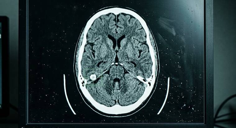
Location, shape and density pointed not to an aneurysm but to a calcification of the right tentorium — a harmless calcified membrane that requires nothing.
You cannot judge the vessels from a study that does not show them. We recommended MRA and a brain MRI — without contrast and without radiation exposure.
There is no aneurysm. The vessels are fine. The tentorium calcification on the right is exactly where we described it. The child is healthy.
“No evidence of aneurysm”
— the wording of our report before further testing
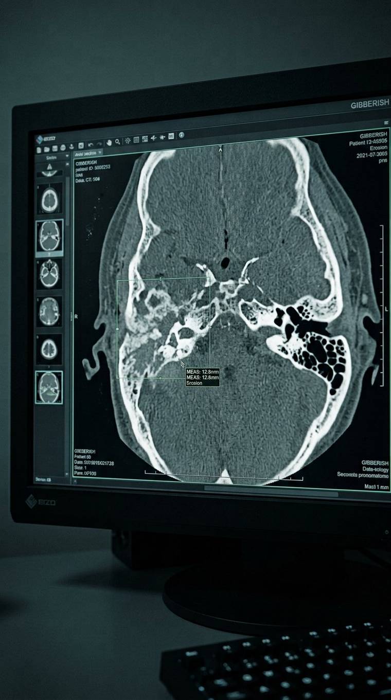
The child was prescribed a long course of antibiotics. The treatment did not help; the condition kept worsening.
We took the temporal bone CT into review and re-read the slices ourselves.
Soft-tissue content with bone destruction — we interpreted the finding as a cholesteatoma.
MRI of the middle ear in DWI — diffusion-weighted images make it possible to distinguish a cholesteatoma from inflammatory content.
MRI confirmed the cholesteatoma. Surgeons operated on the child. His life is no longer in danger; he has recovered.
The antibiotics did not work because they were treating the wrong thing
— a cholesteatoma cannot be treated conservatively, it requires surgery
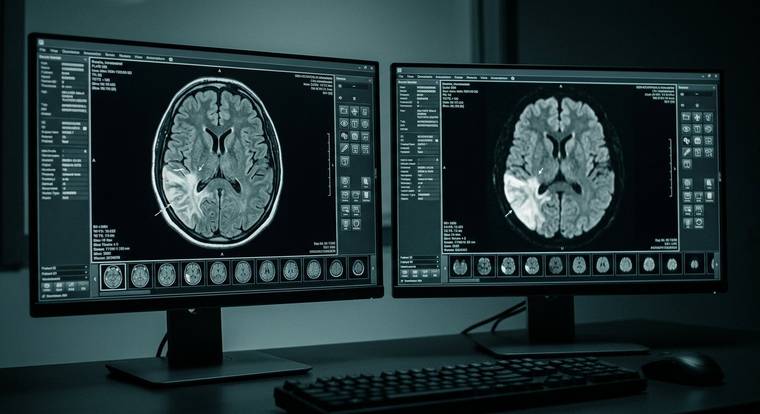
A brain MRI described a glial-series lesion. Surgery was scheduled for the patient.
Our specialists reviewed the study before any intervention.
The location, shape and signal characteristics of the lesion pointed not to a tumor but to post-ischemic changes.
It was a post-ischemic lesion: the patient had had a stroke that went unrecognized in time. Tumor surgery was not needed.
Not a glial tumor, but the aftermath of a stroke
— the difference between neurosurgery and neurological treatment
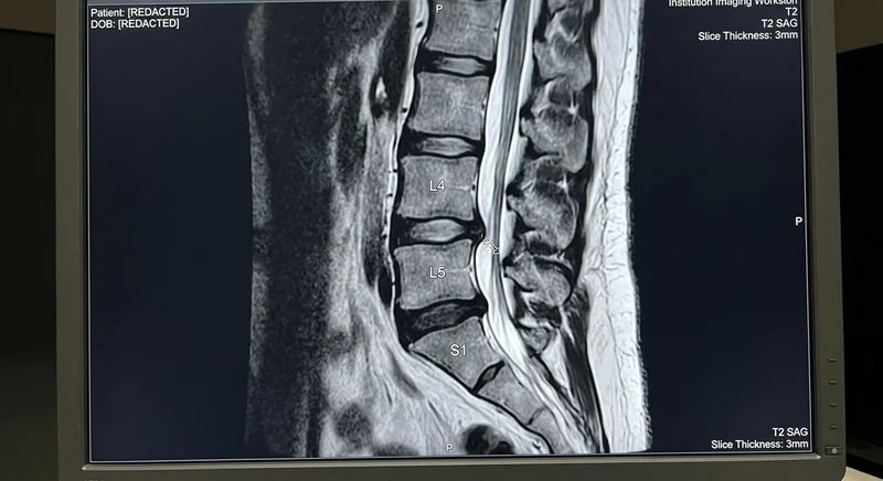
A 42-year-old patient with acute low back pain was diagnosed with a sequestered L5-S1 herniation compressing a nerve root, and urgent surgery (discectomy) was scheduled.
The physician correlated T1, T2 and STIR sequences in all planes: a pronounced edema of the posterior longitudinal ligament had been mistaken for a sequestrum. An ordinary 3.2 mm protrusion without compression of the dural sac or nerve was identified.
The surgery was canceled. A course of conservative therapy and physiotherapy followed. Three weeks later the pain was resolved and the patient returned to work — no incisions, no anesthesia.
Ligament edema instead of a herniated sequestrum
— the difference between open spinal surgery and 3 weeks of physiotherapy
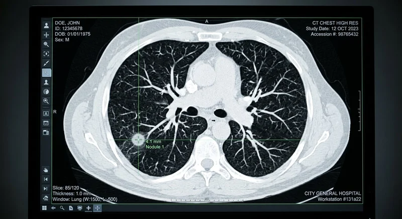
A 56-year-old patient was having a routine chest X-ray. On a plain X-ray, small lesions up to 5 mm in the lung apices are often hidden behind the clavicle and ribs.
The family insisted on a CT and sent the archive for a second opinion. In the lung window, the physician identified a solid 4.2 mm lesion with spiculated contours in segment S1 of the right lung.
Early detection of an in-situ adenocarcinoma before symptoms and metastases. Organ-sparing thoracoscopic resection was performed. The patient is fully healthy, without chemotherapy.
A 4 mm lesion detected before symptoms appeared
— the difference between a complete cure at stage 1 and an advanced cancerAll anatomical zones, tomographic and radionuclide modalities. A separate area of expertise — oncopathology and vascular studies, where the cost of interpretation is highest.
Focal changes, lesions, calcifications. MRA and CT angiography — assessment of vessels, aneurysms and malformations.
BRAIN MRIMRACT ANGIOThyroid, salivary glands, neck lymph nodes. Echogenicity, contour, microcalcifications, category and drainage zones.
NECK CTMRIPET-CTLungs, mediastinum, pleura, breasts. Lesions, nodules, lymph nodes, assessment of disease extent.
CHEST CTMSCTPET-CTLiver, pancreas, kidneys, adrenal glands, lymph nodes. Characterization of lesions and follow-up over time.
CTMRIMSCTOvaries, uterus, cervix, prostate. Septa, solid components, contrast uptake, free fluid.
MRICTPET-CTJoint MRI: ligaments, menisci, cartilage, bone marrow edema. CT and MSCT — bone structure, trauma, focal changes.
JOINT MRICTMSCTScintigraphy and SPECT: skeletal system, thyroid and parathyroid glands, kidneys, myocardium. Assessment of tracer uptake and hyperfixation foci.
SCINTIGRAPHYSPECTSPECT-CTA clear and transparent process, without queues or waiting.
Attach a DICOM archive from your disc in the website form or paste a Yandex.Disk, Google Drive or Mail.ru Cloud link (1–2 minutes).
A physician opens the files on a workstation and confirms that all slices and series are readable before your order is confirmed.
Payment by bank details. A top-category specialist studies your scan slice by slice from scratch, without relying on third-party templates.
An official protocol signed and stamped by the physician. You can ask any follow-up questions about the results in plain language.

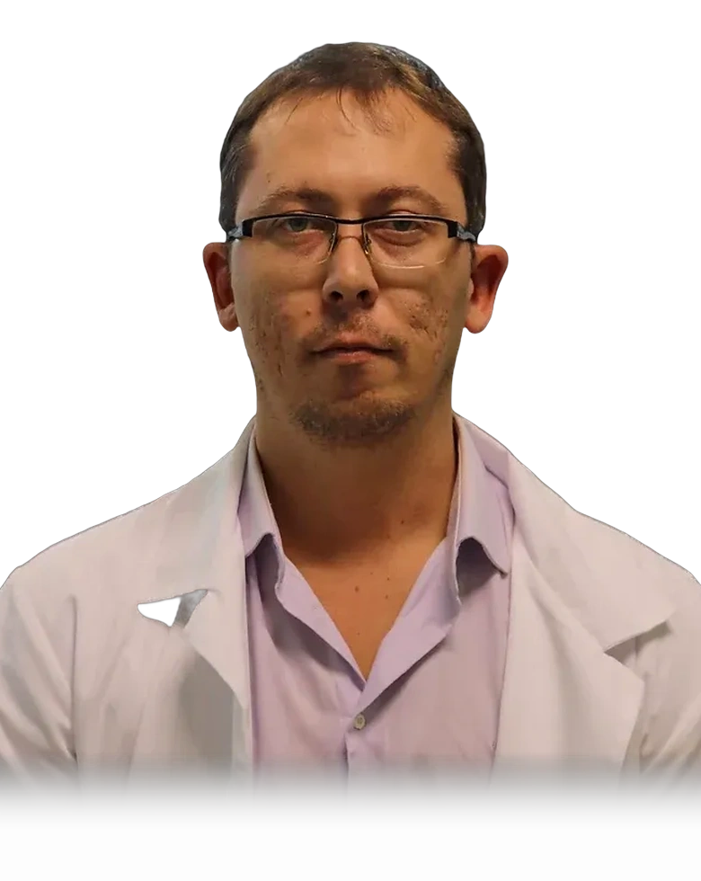
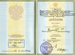
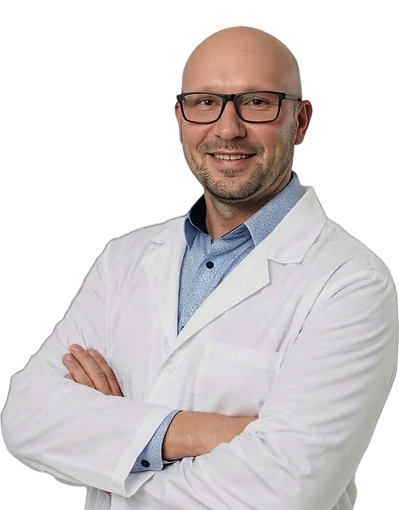
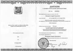
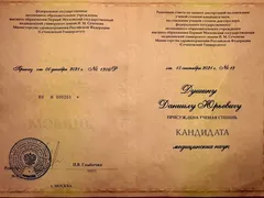
Medical degree

X-ray imaging

PhD
Honest fixed prices from the official price list. No hidden surcharges.
Everything worth knowing before sending your scan for a second opinion.
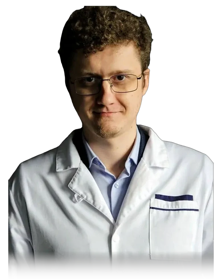
Yes, absolutely. You receive an official written imaging report in an established format with a detailed description of the slices, a conclusion, recommendations and the personal signature of a certified radiologist (MD PhD or top-category physician). With this document you can consult your treating physician, an oncologist, a neurologist, a neurosurgeon or any clinic.
Yes. Archive studies are invaluable for assessing dynamics (has the lesion grown, is the process stable, has the treatment worked). Moreover, if symptoms or pain persist, an independent review of the older slices can reveal hidden details and micro-changes that may have been missed in the first, cursory reading.
By law, a diagnostic center or clinic is obliged to provide a free digital copy of your study on CD/DVD or your USB drive. Simply contact the reception or archive of the clinic where you had your scan and request the DICOM archive.
A standard independent report is ready within 24–48 hours. If a decision about surgery or hospitalization must be made today, an urgent tariff is available — a priority queue within 7 hours.
Yes — we never leave a patient alone with medical jargon. After receiving the report you can ask any follow-up questions in Telegram (@mri_online) or by e-mail, or order a 40-minute video review of your scans.
All data and files are transferred over an encrypted SSL protocol (HTTPS) and stored in a protected environment. We strictly observe medical confidentiality and personal data protection laws.
We need the original DICOM in full, as an archive: the tomograms themselves — not a photo of the film and not the text of the report.
Files up to 20 MB can be attached directly in the form. If your study is larger — upload the archive to Yandex.Disk, Mail.ru Cloud or Google Drive and paste the link.
Not sure how to copy a disc or create a ZIP? Download the illustrated PDF guide in the “Price list and guide” section ➔
Not sure whether all the slices were written to the disc or whether the file will open? Attach an archive or a link — a physician will check readability, series completeness and suitability of the scans for expert review free of charge, before payment.
Don’t want to fill in the form — write directly on Telegram (@mri_online_gr) or by email; the archive will still be needed as a file, not as a photo.
If you are holding a report that frightens you — send us your scan. A second look costs far less than a month of uncertainty.
Send a requestReport within 24 hours · remote, worldwide
The international standard for medical images DICOM is the same for every scanner in the world (Siemens, GE, Philips, Canon, Toshiba). We perform a detailed slice-by-slice review of every image, rule out scanning artifacts, find hidden micro-lesions and issue an official written report with a seal within 24 hours.
Brain, pituitary, neck vessels and the circle of Willis (MRA), spine, joints (menisci, ligaments), pelvis (PI-RADS), abdominal cavity, MRCP.
High-resolution CT (HRCT) of the lungs, mediastinum, bolus contrast enhancement of the abdominal cavity, CT angiography of the aorta and peripheral arteries, temporal bones, sinuses.
Assessment of the coronary bed, coronary calcium index (Agatston), visualization of soft and calcified atherosclerotic plaques, degree of stenosis.
Whole-body PET-CT with 18F-FDG, PSMA, DOPA. Assessment of the primary tumor, detection of micrometastases, TNM staging, follow-up after chemotherapy and radiotherapy.
Largest centers: Moscow, St. Petersburg, Novosibirsk, Yekaterinburg, Kazan, Nizhny Novgorod, Chelyabinsk, Krasnoyarsk, Samara, Ufa, Rostov-on-Don, Omsk, Krasnodar, Voronezh, Perm, Volgograd, Saratov, Tyumen, Tolyatti, Barnaul, Izhevsk, Makhachkala, Khabarovsk, Vladivostok, Irkutsk, Yaroslavl, Kemerovo, Tomsk, Orenburg, Novokuznetsk, Ryazan, Astrakhan, Penza, Lipetsk, Tula, Kirov and all other cities of Russia.
Kazakhstan: Almaty, Astana, Shymkent, Aktobe, Karaganda, Taraz, Pavlodar, Oskemen, Semey, Atyrau, Kostanay, Oral, Aktau.
Belarus: Minsk, Gomel, Mogilev, Vitebsk, Grodno, Brest, Bobruisk, Baranovichi, Borisov, Pinsk, Soligorsk.
CIS: Uzbekistan (Tashkent), Kyrgyzstan (Bishkek), Armenia (Yerevan), Azerbaijan, Georgia.
USA & Canada: New York, Los Angeles, Chicago, Houston, Miami, Toronto, Vancouver, Montreal, Calgary.
UK & Norway: London, Manchester, Birmingham, Edinburgh, Oslo, Bergen, Trondheim, Stavanger.
DACH & Israel: Germany (Berlin, Munich, Frankfurt), Austria (Vienna), Switzerland, Israel (Tel Aviv).
MRI & CT second reading · MRT / CT Zweitmeinung · MR tolkning andre vurdering.

Contact us in whichever way is convenient, or see the service details.